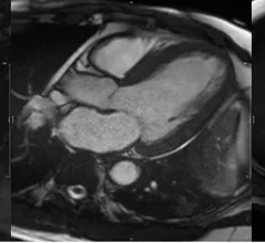
Magnetic Resonance Imaging

Magnetic resonance imaging (MRI) is the gold standard imaging modality for soft tissues. It produces detail cross-sectional images of soft tissue and bone anatomy, including muscles, tendons, ligaments, brain and organs, without the use of ionizing radiation. In addition to orthopedic imaging, MRI is also used for heart, brain and breast. MRI uses gadolinium contrast in many exams to highlight tissues and blood vessels, which enhances images and offers better diagnostic quality. It can also be used in conjunction with PET scans. How does MRI work? MR creates images by using powerful magnets to polarize hydrogen atoms in water (the body is made of of more than 80% water) so they face in one direction. A radiofrequency pulse is then used to ping these atoms, causing them to wobble, or resonate. The MRI coils detect this and computers can assemble images from the signals. Basic MRI scans will focus on the resonance of fat and water in two different sequences, which highlight and contrast different features in the anatomy.

Less experienced radiologists benefit from deep learning models when scouting for intracranial aneurysms

Deep learning models can increase reader accuracy while simultaneously decreasing interpretation times when evaluating imaging for intracranial aneurysms.

Thumbnail

Functional MRI findings could open the door for more accurate ADHD diagnosis and management

Authors of the new study suggested that their findings, once further validated in larger cohorts, could provide objective measures to diagnose ADHD and monitor treatment responses in the future. 

Thumbnail

New study highlights dangers of metallic fabrics in face masks, athletic clothing for MRI patients

The metallic elements in the fabrics are often missing from labels, posing thermal burn risks for MRI patients. 

Thumbnail

Hybrid multidimensional prostate MRI is 'a step in the right direction' for quantitative assessments

Using hybrid multidimensional MRI exams in lieu of multiparametric MRI for the assessment of prostate cancer could reduce interpretation times while increasing interobserver agreement. 

Ischemic stroke CT imaging. Images courtesy of RSNA

CT imaging alone found noninferior to CT/MR combo for acute ischemic stroke patients

“The value of MRI added to CT in patients such as these should not be presumed."

CMR of a patient showing evidence of myocardial hypertrophy

Cardiac MRI findings linked with worse outcomes in hypertension patients

Nonischemic LGE was revealed to be independently associated with worse outcomes in hypertension patients. 

New MRI findings suggest that even moderate drinkers are at risk of alcohol-related cognitive decline

In the largest known study to date on the relationship between moderate alcohol consumption and iron homeostasis, experts suggested that as little as four drinks per week can increase the presence of iron deposits on imaging.

Thumbnail

New MRI method can identify early signs of Parkinson's

The researchers are now working on developing a qMRI method that can be incorporated into clinical settings, with an anticipated timeline of three to five years. 

Around the web

Positron, a New York-based nuclear imaging company, will now provide Upbeat Cardiology Solutions with advanced PET/CT systems and services. 

The nuclear imaging isotope shortage of molybdenum-99 may be over now that the sidelined reactor is restarting. ASNC's president says PET and new SPECT technologies helped cardiac imaging labs better weather the storm.

CMS has more than doubled the CCTA payment rate from $175 to $357.13. The move, expected to have a significant impact on the utilization of cardiac CT, received immediate praise from imaging specialists.