Magnetic Resonance Imaging

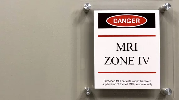
Magnetic resonance imaging (MRI) is the gold standard imaging modality for soft tissues. It produces detail cross-sectional images of soft tissue and bone anatomy, including muscles, tendons, ligaments, brain and organs, without the use of ionizing radiation. In addition to orthopedic imaging, MRI is also used for heart, brain and breast. MRI uses gadolinium contrast in many exams to highlight tissues and blood vessels, which enhances images and offers better diagnostic quality. It can also be used in conjunction with PET scans. How does MRI work? MR creates images by using powerful magnets to polarize hydrogen atoms in water (the body is made of of more than 80% water) so they face in one direction. A radiofrequency pulse is then used to ping these atoms, causing them to wobble, or resonate. The MRI coils detect this and computers can assemble images from the signals. Basic MRI scans will focus on the resonance of fat and water in two different sequences, which highlight and contrast different features in the anatomy.

Cardiac MRI findings can predict incident CVD years before onset

Prior imaging of the descending thoracic and abdominal aorta in study participants revealed thoracic aortic wall area (AWA), plaque prevalence and plaque volumes to be independently associated with incident CVD.

Photo and caption courtesy of American Journal of Roentgenology (AJR), American Roentgen Ray Society (ARRS). Used with permission.

Diffusion-weighted MRI boosts detection of locally recurrent pancreatic cancer

MRI with DWI images showed higher sensitivity in detecting local recurrence after PDAC resection.

Doctor patient with masks

New imaging technique detects post-COVID lung abnormalities

Conventional imaging did not detect the abnormalities, which MRI scans identified up to one year after infection.

Some respiratory face masks are unsafe for MRIs, study finds

Five out of eight filtering face piece (FFP-3) masks studied contained ferromagnetic components.

liver cancer

Measurements from functional MRI scans offer non-invasive boost in liver disease assessment

By using a combination of functional liver imaging scores (FLIS) derived from gadoxetic acid (GA)-enhanced MRI and splenic diameter measured on MRI, the experts were able to non-invasively assess liver impairment.

Thumbnail

New 7T MRI scanners could increase radiologists' role in treating neurological conditions

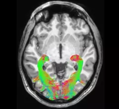
7T MRI brain scans can analyze the locus coeruleusan area difficult to visualize on standard scannersof patients with Parkinson’s and progressive supranuclear palsy to identify those most likely to respond to certain treatment strategies.

Evidence points to optimal MRI sequence for detecting insufficiency fractures

Because sacral insufficiency fractures do not always show a fracture line, they can be difficult to diagnose or even misdiagnosed as bone metastasis, which could result in additional treatments like radio-chemotherapy.

Thumbnail

Prostate cancer recurrence scoring system yields 'promising' results

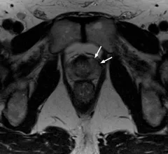
An international panel of experts recently developed and validated a reporting assessment scoring system that analyzes the location and extent of prostate cancer recurrence. 

Around the web

Positron, a New York-based nuclear imaging company, will now provide Upbeat Cardiology Solutions with advanced PET/CT systems and services. 

The nuclear imaging isotope shortage of molybdenum-99 may be over now that the sidelined reactor is restarting. ASNC's president says PET and new SPECT technologies helped cardiac imaging labs better weather the storm.

CMS has more than doubled the CCTA payment rate from $175 to $357.13. The move, expected to have a significant impact on the utilization of cardiac CT, received immediate praise from imaging specialists.