Magnetic Resonance Imaging

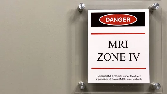
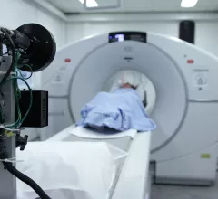
Magnetic resonance imaging (MRI) is the gold standard imaging modality for soft tissues. It produces detail cross-sectional images of soft tissue and bone anatomy, including muscles, tendons, ligaments, brain and organs, without the use of ionizing radiation. In addition to orthopedic imaging, MRI is also used for heart, brain and breast. MRI uses gadolinium contrast in many exams to highlight tissues and blood vessels, which enhances images and offers better diagnostic quality. It can also be used in conjunction with PET scans. How does MRI work? MR creates images by using powerful magnets to polarize hydrogen atoms in water (the body is made of of more than 80% water) so they face in one direction. A radiofrequency pulse is then used to ping these atoms, causing them to wobble, or resonate. The MRI coils detect this and computers can assemble images from the signals. Basic MRI scans will focus on the resonance of fat and water in two different sequences, which highlight and contrast different features in the anatomy.

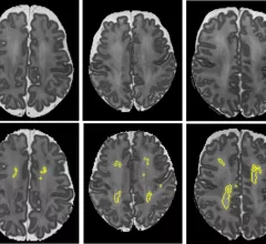
Poor midlife health leads to advanced brain aging, MRI analysis shows

Though the participants’ actual ages ranged from 69 to 72 years old, the experts’ predictive analysis revealed their estimated current brain ages to be between 46 and 93.

idiopathic intracranial hypertension

MRI a useful tool for avoiding invasive procedures for suspected idiopathic intracranial hypertension

The experts maintained that these MRI parameters support the diagnosis of IIH in clinically suspicious cases and could help reduce the use of invasive procedures when aligned with symptoms.

Thumbnail

Machine learning model uses MRI data to identify candidates for liver transplant

When applied to MRI and clinical features, different machine learning models were recently shown to reliably predict post-treatment recurrences of hepatocellular carcinoma. 

Thumbnail

Deep learning system outperforms rads in brain tumor identification and classification

The new findings findings represent “a step toward improved tumor diagnoses," according to authors of a new study published in JAMA.

Placental MRI can predict adverse pregnancy outcomes early on in gestation

A new study highlights the benefits of MRI for determining pregnancy risks related to placental dysfunction, opening doors for earlier care management. 

New MRI algorithm could qualify more epilepsy patients for surgery

When applied to brain imaging, the algorithm assists in the detection of subtle abnormalities in instances of drug-resistant focal cortical dysplasia (FCD), which is known to cause epilepsy and is historically difficult to detect. 

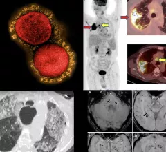
COVID-19 medical imaging examples of various clinical presentations. SARS-CoV-2 clinical imaging presentations.

PHOTO GALLERY: What does COVID-19 look like on medical imaging?

This image gallery shows what the various clinical presentations associated with the COVID-19 virus that have been documented during the coronavirus pandemic.

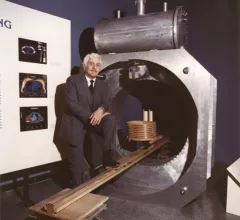
father of #MRI, Raymond Damadian

'Father of MRI' dies at 86

The science community lost a man who pioneered one of medical imaging’s most important developments when Raymond Vahan Damadian passed away August 3. 

Around the web

Positron, a New York-based nuclear imaging company, will now provide Upbeat Cardiology Solutions with advanced PET/CT systems and services. 

The nuclear imaging isotope shortage of molybdenum-99 may be over now that the sidelined reactor is restarting. ASNC's president says PET and new SPECT technologies helped cardiac imaging labs better weather the storm.

CMS has more than doubled the CCTA payment rate from $175 to $357.13. The move, expected to have a significant impact on the utilization of cardiac CT, received immediate praise from imaging specialists.