Breast Imaging

Breast imaging includes imaging modalities used for breast cancer screenings and planning therapy once cancer is detected. Mammography is the primary modality used. Mammogram technology is moving from 2D full-field digital mammography (FFDM) to breast tomosynthesis, or 3D mammography, which helps reduce false positive exams by allowing radiologists to look through the layers of tissue. Overlapping areas of dense breast tissue on 2D mammograms appear similar to cancers and 3D tomo helps determine if suspect areas are cancer or not. About 50% of women have dense breast tissue, which appears white on mammograms, the same as cancers, making diagnosis difficult. Radiologists use the Breast Imaging Reporting and Data System (BI-RADS) scoring system to define the density of breast tissue. Many states now require patients to be notified if they have dense breasts so they understand their mammograms might be suboptimal and they should use supplemental imaging that can see through the dense areas. This includes tomosythesis, breast ultrasound, automated breast ultrasound (ABUS), breast MRI, contrast enhanced mammography and nuclear imaging, including positron emission mammography (PEM).

axillary lymphadenopathy after COVID booster

Follow-up imaging for reactive adenopathy s/p COVID booster might not be necessary

Experts recently gained a better understanding of how axillary lymphadenopathy presents on breast MRI following COVID boosters.

Thumbnail

Is it OK to delay follow-ups for 'probably benign' abnormalities on mammography screening?

Many were concerned that delays in care inflicted by COVID restrictions also would potentially delay diagnoses, causing undue harm for patients.

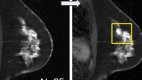
synthetic contrast-enhanced breast MRI

GBCA dose drops significantly in breast MRI thanks to machine learning

The use of synthetic images could reduce the amount of gadolinium-based contrast agents needed for breast MRI examinations, according to new data published this week in Radiology

#covidboostersideeffects #axillarylympadenopathy

COVID booster side effects: How long does axillary lymphadenopathy last after vaccination?

Axillary lymphadenopathy following COVID vaccination and/or boosters is a finding all radiologists must be mindful of when interpreting imaging, but new data clarify the timeline for when the side effect should resolve.

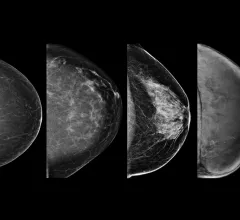
Example of the four types of breast tissue density. The density of fibroglandular tissue inside the breast impacts the ability to easily see cancers. Cancers are very easy to spot in fatty breasts, but are almost impossible to find in extremely dense breasts. These examples show craniocaudal mammogram findings characterized as almost entirely fatty (far left), scattered areas of fibroglandular density (second from left), heterogeneously dense (second from right), and extremely dense (far right). RSNA

How do hormonal contraceptives affect breast density?

How recent developments in hormonal contraceptives affect breast density is an important consideration, as an increase in density category increases cancer risk.

Thumbnail

AI competition furthers research on computer-aided detection in breast imaging

For the challenge, eight teams were tasked with developing algorithms capable of achieving high sensitivity for lesion detection on DBT exams.

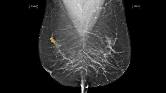
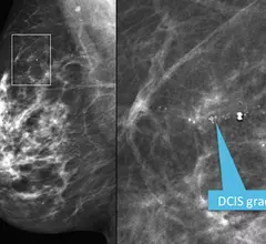
Mediolateral oblique view from a screening mammogram in a 54-year-old woman shows a small cluster of microcalcifications in the upper outer quadrant of the right breast. The right image shows a detailed spot magnification view of the calcifications. Stereotactic biopsy revealed grade 2 ductal carcinoma in situ (DCIS). RSNA image. Image of breast micro calcifications associated with breast cancer.

Why the way microcalcifications on mammograms are regarded could change

New research suggests that the tissue environment where microcalcifications of the breast are formed could hold clues into how breast cancer progresses.

#DCIS #invasivebreastcancer #cancerupgrade #breastMRI

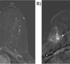
Ultrafast MRI predicts breast cancer upgrade

Preoperative ultrafast MRI could help guide biopsy and surgical management decision in patients with ductal carcinoma in situ. 

Around the web

The nuclear imaging isotope shortage of molybdenum-99 may be over now that the sidelined reactor is restarting. ASNC's president says PET and new SPECT technologies helped cardiac imaging labs better weather the storm.

CMS has more than doubled the CCTA payment rate from $175 to $357.13. The move, expected to have a significant impact on the utilization of cardiac CT, received immediate praise from imaging specialists.

The newly cleared offering, AutoChamber, was designed with opportunistic screening in mind. It can evaluate many different kinds of CT images, including those originally gathered to screen patients for lung cancer. 

Trimed Popup
Trimed Popup