Breast Imaging

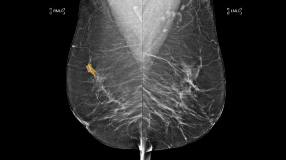
Breast imaging includes imaging modalities used for breast cancer screenings and planning therapy once cancer is detected. Mammography is the primary modality used. Mammogram technology is moving from 2D full-field digital mammography (FFDM) to breast tomosynthesis, or 3D mammography, which helps reduce false positive exams by allowing radiologists to look through the layers of tissue. Overlapping areas of dense breast tissue on 2D mammograms appear similar to cancers and 3D tomo helps determine if suspect areas are cancer or not. About 50% of women have dense breast tissue, which appears white on mammograms, the same as cancers, making diagnosis difficult. Radiologists use the Breast Imaging Reporting and Data System (BI-RADS) scoring system to define the density of breast tissue. Many states now require patients to be notified if they have dense breasts so they understand their mammograms might be suboptimal and they should use supplemental imaging that can see through the dense areas. This includes tomosythesis, breast ultrasound, automated breast ultrasound (ABUS), breast MRI, contrast enhanced mammography and nuclear imaging, including positron emission mammography (PEM).

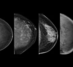
Example of the four types of breast tissue density. The density of fibroglandular tissue inside the breast impacts the ability to easily see cancers. Cancers are very easy to spot in fatty breasts, but are almost impossible to find in extremely dense breasts. These examples show craniocaudal mammogram findings characterized as almost entirely fatty (far left), scattered areas of fibroglandular density (second from left), heterogeneously dense (second from right), and extremely dense (far right). RSNA

Experts developed a deep learning model that can estimate breast density

When tested, the model achieved a performance comparable to that of human experts.

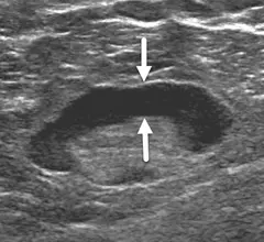
Is scanning the axilla during diagnostic breast ultrasound necessary?

Experts recently questioned the necessity of scanning the axilla region during diagnostic breast ultrasound, as new data indicate that it is minimally beneficial for cancer detection. 

Thumbnail

Follow-up adherence affected by how and when imaging orders are placed

These are factors that healthcare systems can and should control, experts recently suggested in a new JACR paper.

Chat GPT

ChatGPT offers 'pretty amazing' recommendations on breast cancer screening, but oversight remains critical

A team of experts with the University of Maryland School of Medicine recently presented ChatGPT with a set of questions relative to breast cancer screening recommendations to determine whether the program could reliably offer appropriate guidance.

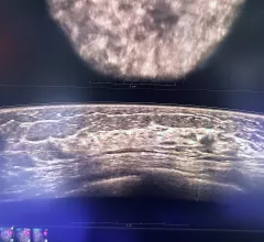
Automated breast ultrasound (ABUS) allows for reproducible breast imaging without variation based on which sonographer performs the exam. It also can help centers were they are short on qualified breast sonographers.  Breast ultrasound can help identify cancers, or benign cysts, even in women with very dense breast tissue. At the GE Healthcare booth at RSNA.

Automated ultrasound excels for women with dense breasts

Could automated breast ultrasound replace handheld ultrasound?

Example of the four types of breast tissue density. The density of fibroglandular tissue inside the breast impacts the ability to easily see cancers. Cancers are very easy to spot in fatty breasts, but are almost impossible to find in extremely dense breasts. These examples show craniocaudal mammogram findings characterized as almost entirely fatty (far left), scattered areas of fibroglandular density (second from left), heterogeneously dense (second from right), and extremely dense (far right). RSNA

Breast density notification laws: How will the FDA's updates affect radiologists and referring providers?

The FDA will soon require mammography providers to inform all patients of their breast density status. To anticipate how the update might affect key stakeholders, it’s important to understand exactly what has changed. 

axillary lymphadenopathy after COVID booster

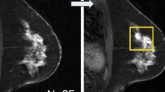
Follow-up imaging for reactive adenopathy s/p COVID booster might not be necessary

Experts recently gained a better understanding of how axillary lymphadenopathy presents on breast MRI following COVID boosters.

Thumbnail

Is it OK to delay follow-ups for 'probably benign' abnormalities on mammography screening?

Many were concerned that delays in care inflicted by COVID restrictions also would potentially delay diagnoses, causing undue harm for patients.

Around the web

CMS has more than doubled the CCTA payment rate from $175 to $357.13. The move, expected to have a significant impact on the utilization of cardiac CT, received immediate praise from imaging specialists.

The newly cleared offering, AutoChamber, was designed with opportunistic screening in mind. It can evaluate many different kinds of CT images, including those originally gathered to screen patients for lung cancer. 

AI-enabled coronary plaque assessments deliver significant value, according to late-breaking data presented at TCT. These AI platforms have gained considerable momentum in recent months, receiving expanded Medicare coverage in addition to a new Category I CPT code.

Trimed Popup
Trimed Popup