Breast Imaging

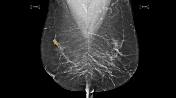
Breast imaging includes imaging modalities used for breast cancer screenings and planning therapy once cancer is detected. Mammography is the primary modality used. Mammogram technology is moving from 2D full-field digital mammography (FFDM) to breast tomosynthesis, or 3D mammography, which helps reduce false positive exams by allowing radiologists to look through the layers of tissue. Overlapping areas of dense breast tissue on 2D mammograms appear similar to cancers and 3D tomo helps determine if suspect areas are cancer or not. About 50% of women have dense breast tissue, which appears white on mammograms, the same as cancers, making diagnosis difficult. Radiologists use the Breast Imaging Reporting and Data System (BI-RADS) scoring system to define the density of breast tissue. Many states now require patients to be notified if they have dense breasts so they understand their mammograms might be suboptimal and they should use supplemental imaging that can see through the dense areas. This includes tomosythesis, breast ultrasound, automated breast ultrasound (ABUS), breast MRI, contrast enhanced mammography and nuclear imaging, including positron emission mammography (PEM).

Thumbnail

Why breast MRI may be helpful to male patients, too

For the last 10 years, researchers from the University of Texas MD Anderson Cancer Center have been compiling examples of when breast MRI may be helpful for male patients, according to a study published online Jan. 10 in Diagnostic Radiology.  

Thumbnail

Fewer repeat surgeries in women imaged pre-op with breast MRI

While preoperative breast MRI rarely changes prior decisions to perform mastectomy on women with biopsy-proven breast cancer, the extra imaging can reduce the odds of needing a second trip under the knife for patients undergoing breast-conserving surgery.

Thumbnail

Researchers call for making breast MRI a standalone screening exam

Annual screening mammography adds no value to women who are high risk for breast cancer and, as a matter of course, are already getting screened each year with breast MRI, according to a study conducted at the University of Toronto and published online June 6 in Radiology.

Thumbnail

‘Controversial’ preoperative breast MRI more accurate than mammography/ultrasound for many patients

A third of breast-cancer patients undergoing preoperative mammography and ultrasound would be more accurately imaged for tumor size with breast MRI, according to a study published online April 13 in the Journal of Surgical Oncology. 

Thumbnail

Affordable breast MRI protocol improving detection in women with dense breasts

Via Radiology, based in Seattle, is the first in the Pacific Northwest to offer a new type of MRI screening for breast cancer patients.

Thumbnail

Danish study challenges value of screening mammography, draws sharp criticism from U.S. orgs

European researchers have reviewed a huge mammography dataset and found no association between screening mammography and a reduction in the incidence of advanced breast cancer. 

Thumbnail

Expanded genetic testing points more women to screening breast MRI

Testing for gene mutations beyond just BRCA1 and BRCA2 would indicate screening breast MRI and other proactive measures for many women who would not have been considered candidates for such measures going by family history alone, according to a large-sample study published online Dec. 21 in the Journal of the American College of Radiology.

Thumbnail

For high-risk women, even small lesions on breast MRI call for prompt assessment and biopsy

A U.K. study based on a departmental audit has confirmed previous research suggesting that MRI-detected small enhancing masses and new small enhancing foci, including those smaller than 5 millimeters, should be considered suspicious in women at high risk for breast cancer.

Around the web

Positron, a New York-based nuclear imaging company, will now provide Upbeat Cardiology Solutions with advanced PET/CT systems and services. 

The nuclear imaging isotope shortage of molybdenum-99 may be over now that the sidelined reactor is restarting. ASNC's president says PET and new SPECT technologies helped cardiac imaging labs better weather the storm.

CMS has more than doubled the CCTA payment rate from $175 to $357.13. The move, expected to have a significant impact on the utilization of cardiac CT, received immediate praise from imaging specialists.