Magnetic Resonance Imaging

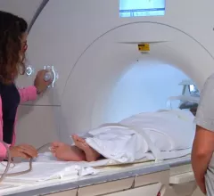
Magnetic resonance imaging (MRI) is the gold standard imaging modality for soft tissues. It produces detail cross-sectional images of soft tissue and bone anatomy, including muscles, tendons, ligaments, brain and organs, without the use of ionizing radiation. In addition to orthopedic imaging, MRI is also used for heart, brain and breast. MRI uses gadolinium contrast in many exams to highlight tissues and blood vessels, which enhances images and offers better diagnostic quality. It can also be used in conjunction with PET scans. How does MRI work? MR creates images by using powerful magnets to polarize hydrogen atoms in water (the body is made of of more than 80% water) so they face in one direction. A radiofrequency pulse is then used to ping these atoms, causing them to wobble, or resonate. The MRI coils detect this and computers can assemble images from the signals. Basic MRI scans will focus on the resonance of fat and water in two different sequences, which highlight and contrast different features in the anatomy.

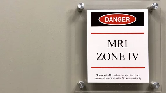
The American College of Radiology is seeking input on new MRI safety guidelines

The most recent updates contain “substantial new content and safety recommendations," the ACR reported recently. 

Thumbnail

New neural-redundancy metric accurately detects mild cognitive impairment

Redundant brain pathways can serve as pre-symptomatic indicators of mild cognitive impairment (MCI) in aging individuals, allowing care teams to design first-line neuropsychological exercises for slowing the possible approach of Alzheimer’s disease.

Thumbnail

New scoring system predicts rupture risk for patients with arteriovenous malformations

The risk stratification system was put to the test in more than 3,000 cases and proved itself to be a beneficial tool in predicting future AVM rupture across multiple risk groups.

kid child pediatric MRI imaging

New data highlight inconsistencies in imaging protocols for pediatric appendicitis

Utilization of MRI for cases of suspected appendicitis in the pediatric population is beneficial, yet its use varies across institutions.

Thumbnail

Deep learning model predicts Alzheimer's using routine MRI exams

When put to the test, the new model was able to predict Alzheimer’s risk with 90.2% accuracy.

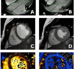
Experts develop cardiac MRI reference values for healthy adolescents

The newly developed reference values could have “direct implications for clinical practice” and can be used by any provider to determine whether an adolescent falls within the normal range of measurements for their age group.

Thumbnail

GE HealthCare's new MRI contrast agent advances to clinical trial

Experts are hopeful that the first-of-its-kind injectable manganese-based macrocyclic agent will offer a suitable alternative to GBCAs, as it can be more efficiently eliminated from the body.

Thumbnail

MRI findings differ significantly in boys and girls who have sustained concussions

Findings from functional MRI brain scans following concussions in pediatric patients indicate that injury recovery times may differ between sexes. 

Around the web

Positron, a New York-based nuclear imaging company, will now provide Upbeat Cardiology Solutions with advanced PET/CT systems and services. 

The nuclear imaging isotope shortage of molybdenum-99 may be over now that the sidelined reactor is restarting. ASNC's president says PET and new SPECT technologies helped cardiac imaging labs better weather the storm.

CMS has more than doubled the CCTA payment rate from $175 to $357.13. The move, expected to have a significant impact on the utilization of cardiac CT, received immediate praise from imaging specialists.

Trimed Popup
Trimed Popup